.png)
Ultrasound-Guided Tenotomy
Breaking up the calcium that's stuck in your tendon
Ultrasound-guided tenotomy uses a needle to clear calcium deposits and damaged tissue out of a tendon and restart the healing that stalled.
It’s done in the office, under local anesthetic, and it ends with a bandage over the entry point. No incision, no stitches.
What is tenotomy?
When there's calcium in a tendon, or a stretch of it has degenerated and stayed that way, the usual treatments are aimed at changing how the tendon behaves rather than what's inside it.
Therapy loads it so it remodels, cortisone quiets inflammation that often wasn't the problem to begin with, and even an injection like PRP can’t remove hardened calcium deposits. None of them physically affect the material that's actually sitting in there.
Tenotomy does. In plain terms, it's the use of a needle to break up damaged tissue inside a tendon, repeatedly and in a controlled way, in the area that's genuinely degenerated. It clears out the disorganized collagen that healed badly or half healed, and at the same time, it deliberately aggravates the area to kickstart a healing response the tendon had given up on.
That second part is PRP's job description, too. Both treatments are attempts to harness your body's own innate healing capacity and give the healing response a second chance to get it right, with tenotomy getting there by debriding the damaged area and PRP by delivering concentrated healing factors into it.
In tenotomy, there are two needles. The first is the outer needle, which is 18-gauge, or about the size of the needle used for an IV line. Inside it sits a second, smaller needle that oscillates back and forth ten to thirty times a second, breaking up whatever is in front of it: damaged tendon tissue, calcium deposits, or both. The closest everyday comparison is the ultrasonic instrument a dental hygienist uses on hardened plaque, or an electric toothbrush. Rapid oscillation is what makes short work of hardened deposits, and calcium in a tendon is the same kind of problem as plaque on a tooth.
Once those deposits are broken apart, we rinse the area with sterile saline and suction the calcium debris back out through the same needle. What’s left behind is a clean pocket where the diseased tissue used to be, which we can then fill with PRP.
Not quite surgery, and more than an injection
It’s not surgery, and there’s no incision. Everything happens through a single needle entry point the size of an IV stick, and it’s closed at the end with a bandage. No sutures, no steri-strips.
It’s not an injection, either. When people picture an injection, they picture a needle going in, medicine going in, and the needle coming out. This is different, because the needle is being used as an instrument rather than a delivery vehicle. It’s removing tissue, not adding it.
If you’ve had dry needling in physical therapy, you’ve already met the underlying idea: a needle used to provoke a tendon into responding. What’s different here is a matter of degree and precision. Dry needling uses a single fine needle placed by feel. This uses a large-bore needle guided by ultrasound, with an oscillating tip, aimed at tissue we can see on a screen and have confirmed is damaged.
What conditions is tenotomy used for?
The honest answer starts with what tenotomy is capable of, and then narrows to how we actually use it here, because those two things aren’t the same.
Tenotomy has been used for degenerative tendon conditions generally: tennis elbow, gluteal tendinopathy, and the rest of the family of tendon problems that were misdiagnosed for years as inflammatory conditions. It was one of the promising options for those conditions before PRP gained momentum. There are doctors who think tenotomy achieves the same outcomes as PRP for a tendon like that, but Dr. Kiok is not so sure.
Instead, here’s where he lands: for a tendon that’s simply degenerated, with no calcium in it, he goes for PRP first and only. Our patients get consistently strong results with it, and he’s not interested in putting a second procedure head-to-head against something that’s already working.
Where Dr. Kiok reaches for tenotomy is calcium.
Why calcium is a different problem
Calcium deposits anywhere in the body usually mean the same thing: an old internal scar somewhere the body attempted to heal an area and partly got there and partly didn’t. Think of them as small pebbles somewhere you don’t want pebbles, like inside your shoe. They’re abrasive against the tendon they’re sitting in, they can be genuinely painful, and in a highly mobile joint like the shoulder, a large enough deposit can physically stop your arm from reaching the top of its range.
That’s a mechanical problem, and a mechanical problem needs a mechanical solution. PRP will not dissolve calcium, no matter how good the PRP is.
Calcium also rarely turns up on its own. It’s usually there because something substantial happened to that tendon once and it never fully healed, so in practice we tend to find calcium and a degenerated tendon together, in the same place, in the same patient. That combination is the typical picture, and it shapes what we recommend.
The tendon we see this in the most is the shoulder, where calcific tendinopathy can build deposits large enough to block movement outright, but the same logic applies anywhere calcium accumulates, including the Achilles, the patellar tendon, and the elbow. Any tendon carrying too much calcium is one we consider treating this way.
One honest caveat, since this is new to OASIS: as we do more of these, we expect to either widen or narrow where this is offered. Dr. Kiok would rather tell you that now than pretend the list is settled.
How would you know whether your own tendon has calcium in it?
We look at it live, in motion, on a musculoskeletal ultrasound. It’s not a subtle finding you have to hunt for, and it’s not something we have to guess at from your symptoms. We can put a probe on the area during your consultation and tell you then and there.
But finding calcium isn’t automatically a reason to remove it. Plenty of tendons have calcium speckled through them that isn’t restricting anything and isn’t the source of the pain. If a scan shows deposits but we’re not convinced they’re causing a mechanical restriction, Dr. Kiok will say so, and talk through the options rather than defaulting to the needle.
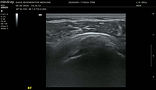
Circled in blue, you can see calcium in the long axis. The bright band is a calcium deposit; the dark shadow beneath it is the ultrasound being completely blocked as the normal bone contour on either side disappears underneath. This is the deposit that was preventing the patient from lifting their arm overhead.

How do we decide who's a candidate?
As with everything we offer, an accurate diagnosis is the non-negotiable foundation. Before we discuss any treatment, we have to be confident about what we’re treating and where it is.
For this procedure, the decision comes down to two questions we answer with ultrasound in the office.
Is there a mechanical problem? That means calcium that’s either physically impinging on movement or causing pain in its own right. In a shoulder, this is usually unmistakable. The humerus gets stuck partway through horizontal abduction or forward flexion, and you can watch the surrounding muscles fire in odd compensatory patterns trying to find a way around the calcification. For a shoulder, a mechanical block like that is the reason we offer this at all.
What’s the state of the tendon around it? A calcium deposit in an otherwise healthy tendon is a purely mechanical problem with a purely mechanical solution. A calcium deposit sitting in a degenerated tendon is two problems, and it takes two treatments.
What the scan shows
Calcium that’s blocking movement or causing pain, with healthy tendon around, above, and below it.
A degenerated tendon with no meaningful calcium.
Calcium and tendon degeneration together, which is the common picture.
What we offer
Tenotomy on its own. There’s no biological problem attached, so there’s nothing for PRP to do.
PRP on its own. This is the bread and butter here, and tenotomy doesn’t improve on it.
The combination. Clear the calcium out, rinse the debris, then backfill the pocket with PRP.
What has to have been tried first?
Therapy and progressive loading, at minimum. For some people, that’s genuinely enough, and if it is, you’re all done. There’s no reason to put a needle in you when loading the tendon properly would have sorted it.
We don’t tend to see people at the beginning of an injury. They come when they’re at their wits’ end, and that shapes what we recommend. When someone is already at that point, and there’s a chunk of calcium in the way, we’re not inclined to offer a half measure. Dr. Kiok would rather pick the most definitive thing he knows of to make the problem go away.
What changes the conversation?
People reasonably assume that a mechanical procedure sidesteps the biology, so that someone who can’t have PRP because of a personal health factor could have this instead. That isn’t how it works. The reasoning is that, in addition to clearing out damaged tissue, tenotomy deliberately aggravates the area to try to restart a healing response, which is PRP’s job description, too. If your body can’t mount that response, neither treatment has much to work with.
So the same host factors limit both: badly controlled diabetes, smoking, malnutrition, etc. If something would disqualify you from PRP, it will probably blunt your response to tenotomy as well.
The good news is that most of those factors are workable. If your body isn’t in a position to heal well today, that doesn’t mean it can’t be in two or three months under our care. OASIS runs its own GLP-1 program for exactly this reason, so getting the metabolic picture right becomes part of the same plan with the same physician rather than a referral somewhere else.
One structural limit worth naming up front: because a rapidly oscillating needle is being used inside a tendon, Dr. Kiok is cautious about offering this to anyone with a substantial partial-thickness tear on top of the degeneration. With a partial tear that’s a third of the tendon’s thickness or less, he’s comfortable proceeding. Anything beyond that and he’ll stay away from tenotomy for now. That isn’t a no; it just usually means reversing the order, treating the tendon with PRP first, and revisiting the calcium at three or six months if it’s still symptomatic, once the tendon underneath has had some healing done.
One more thing belongs in this conversation, and it's about your calendar rather than your tendon. Because rehabilitation after this isn't optional, someone facing months of heavy travel or other interruptions may be better served by delaying the procedure until they can actually do the work. Dr. Kiok would rather have this conversation honestly before he books anything than treat you at a moment when you can’t follow through.
So candidacy here isn’t a yes pile and a no pile. Instead, it rests on what the scan shows, what the tendon around it can tolerate, what your body is currently able to supply, and whether this is the right season of your life to commit to the recovery. The consultation exists to find out how close you are and what it would take to get you the result you’re after.
How does tenotomy compare to the alternatives?
Tenotomy vs. PRP on its own
PRP won’t dissolve calcium, so if a deposit is extensive enough to be part of what’s driving your pain or limiting your movement, no amount of PRP will clear it. That’s not what PRP does.
The reverse is also true: Tenotomy clears diseased tissue and irritates the area enough to restart a healing response, but it doesn’t supply anything to heal with.
The analogy Dr. Kiok uses with patients is a pothole in a road. Tenotomy is a crew turning up and putting cones and flags around the pothole so everyone knows it’s there. Sometimes the attention is enough to get things moving. Alternatively, PRP sends the crew, and the asphalt truck, and the paver. If all we do is irritate an area and the body doesn’t mount a robust enough response on its own, it can settle straight back into the same half-healed state it was already in.
Which is why, when there’s calcium sitting in a tendon that’s also degenerated, the combination is what we’d offer: clear out the calcification debris, suction and rinse the fragments out with sterile saline, then backfill the pocket with PRP. That combination is likely to produce a more robust and durable result than either one on its own. The residual pocket is part of why, though it isn’t the whole reason. The bigger reason is that we’ve done both jobs, the mechanical one and the biological one, in a single visit.
Tenotomy vs. cortisone
Cortisone is a powerful anti-inflammatory, and it’s what most people with a bad tendon have already been offered. The problem is a mismatch: calcific and degenerative tendon disorders aren’t inflammatory conditions. Cortisone is very good at switching off inflammation, and it’s been used for years on problems that don’t have much inflammation to switch off. It’s the wrong tool for this particular job, not because it’s a bad drug, but because it’s aimed at something your tendon isn’t doing.
Tenotomy vs. therapy and loading
If therapy and progressive loading are enough, use them. Tendons respond to load, and for a meaningful number of people, a well-run loading program resolves the problem. Dr. Kiok has no interest in putting a needle into someone who didn’t need one.
Tenotomy vs. surgery
Surgery for degenerative tendon disorders has a poor track record, and the surgeons themselves will usually tell you so. When you hear a surgeon being reluctant to operate on a tennis elbow, that’s not them withholding something from you. That’s an ethical surgeon, because they know that even when they open the area up, debride the damaged tendon, and try to restart a healing response, the damage done on the way in tends to outweigh whatever healing they get on the way out.
Tenotomy is doing something structurally similar to what that surgery is doing, without the incision, the general anesthesia, the sutures, or the recovery that comes with it.
Tenotomy vs. shockwave
Shockwave is a real, non-invasive option for calcific tendon problems. What looks like a massage gun is held over the area for ten or fifteen minutes, and the idea is that it breaks up the calcium from outside the body without a needle ever going in. For someone earlier in the process, that’s a reasonable thing to try, and it has a decent chance of working.
But the downsides are real, too. You need many sessions, it hurts, and it still might not work. Dr. Kiok had shockwave done on his own elbow, for the bone spur and the early tennis elbow that was forming there, and he’ll tell you plainly: it hurt, and it didn’t work for him. That’s one person’s experience and not evidence, but it’s his honest answer when patients ask.
So the decision usually comes down to time and tolerance. If you have several weeks to give it and you’d rather stay non-invasive, shockwave first is a defensible choice. If you don’t have that time, or you’ve already been through it, or you simply want this dealt with definitively, that’s where tenotomy and PRP come in.
Approach
Therapy and progressive loading
Cortisone
Shockwave
PRP
Tenotomy
Surgery
Its strengths
Loading the tendon so it heals in the right direction. Enough on its own for some people.
Switching off inflammation.
Breaking up calcium from outside the body, with nothing entering it.
Supplying what a degenerated tendon needs to finish healing.
Physically removing calcium and damaged tissue, and restarting a stalled healing response.
Reconstructing tissue that’s completely disrupted.
Its limits
Won’t clear a calcium deposit, and won’t rescue a tendon that’s too far gone.
Calcific and degenerative tendon problems aren’t inflammatory, so it’s aimed at the wrong target.
Many sessions, it’s painful, and it doesn’t always work.
Won’t dissolve calcium, and depends on what your body can supply.
Doesn’t supply healing materials on its own. Limited by substantial partial-thickness tears.
For degenerative tendon problems, the damage on the way in tends to outweigh the healing on the way out.
Ready for an answer?
%20(LinkedIn%20Single%20Image%20Ad).png)
Request a consultation with Dr. Kiok to find out if you're a candidate for tenotomy.
What does treatment look like?
The appointment runs a lot like a PRP visit, which surprises most people given how the procedure sounds when it’s described.
We start by numbing the area as thoroughly as we can with local anesthetic infiltrated into the site. If local infiltration isn’t going to be enough on its own, we can add a nerve block higher up the limb.
Not everyone needs one, but for a tennis elbow with a lot of calcification, for example, that would need a radial nerve block further up the arm. A nerve block can affect the motor nerves as well as the sensory ones, so you’d leave in a sling and expect limited use of that wrist for the next few hours.
From there, we bring the area up on ultrasound, identify exactly where we’re working, mark our spots, and run the safety checks. Then we get in there and start debriding. The debridement itself takes around five minutes. After that, we’re either finished, or we backfill the area with PRP. Then a bandage goes over the entry point, and you’re done. No sutures, no stitches, no steri-strips.

How debriding looks under ultrasound.
Afterward
You’re going to be sore. We want to be straightforward about that, because we’ve just gone in and cleared a quantity of material out of your tendon. Expect the elbow or knee or shoulder to be considerably more sore than it would be after a standard PRP injection, and expect that to last a few days.
You’ll be able to drive. Whether you can lift, or want to, is a different question. You can do what you can tolerate, and you probably won’t tolerate much at first. Gripping, lifting weights, or anything like flipping a pan while you cook is not something we’d be optimistic about in the first few days.
After that, the arc looks a lot like PRP. Meaningful improvement typically begins inside the first one to two weeks. It becomes robust by four to six weeks. It reaches its peak at around three months, and the benefit then holds through longer-term follow-up.
Rehabilitation
Rehabilitation is part of every tenotomy after-care plan. Clearing the calcium out addresses what was physically in the way, a structured rehabilitation program is what tells the tendon how to rebuild once it’s gone, and the two together are what produce a durable result rather than a temporary one.
How we know it worked
We don’t rely on how you feel about it in the moment. We look for your pain going down, objective measures improving, and your ability to resume activities you couldn’t do before.
Which measures depends on where we treated. For an elbow, that means strength testing such as grip and wrist extension. For a shoulder, it means range of motion (or pain-free range of motion, where the block was subtler), because getting the arm moving again is the whole point of clearing the deposit that was stopping it.
Tenotomy tends to be a one-and-done procedure. Unless we failed to clear all the calcium, or we had to stop early and leave some behind, we don’t routinely offer a second pass. If the result falls short of what we were after, the more likely next step is a PRP booster rather than going back in with the needle.
Step 1: Consultation and diagnosis
Interview, exam, and in-office ultrasound to establish what’s in the tendon and what state the tendon is in. One hour, $500.
Step 2: Optimization (if needed)
Address anything limiting your ability to heal before we treat.
Step 3: Day of the procedure
Minimal local anesthetic, or a nerve block if the work is extensive. Ultrasound mapping and safety checks.
Step 4: The debridement
Around five minutes of ultrasound-guided debridement, the calcium debris rinsed and suctioned out, and PRP backfilled if that’s the plan.
Step 5: Aftercare and follow-up
A bandage, no stitches. Sore for a few days. Improvement begins in the first one to two weeks, becomes robust by four to six, and peaks at around three months, paired with a structured rehabilitation program throughout.
What are the risks?
Compared to a straightforward injection, this is a more traumatic procedure, and it should be described that way. There’s a needle oscillating rapidly inside your tendon, and it’s removing tissue rather than adding it.
The theoretical risk follows directly from that. If someone aimed this device at a stretch of healthy tendon, they could do real damage without fixing anything. That’s the entire reason it’s done under ultrasound. There’s no guessing about where the needle is, at any point, and Dr. Kiok is only removing tissue he can confirm on the screen is damaged. Calcium in particular is unmistakable on ultrasound, which makes the target about as clear as targets get.
For context on the margin involved: the device manufacturers put the figure at ten to fifteen minutes of deliberate cutting into healthy tissue before you’d expect a tendon to rupture, against roughly five minutes of targeted debridement in an actual procedure. You’d have to be looking for trouble to cause it. That’s a manufacturer’s figure rather than an independent one, and we’d treat it as an order of magnitude rather than a promise, but it tells you where the margin sits.
Because that risk exists at all, this isn’t a procedure we offer to everyone. As above, a substantial partial-thickness tear on a background of degeneration is where we stop. Our line is around a third of the tendon’s thickness, and past that, we’d treat with PRP first and revisit the calcium later.
Beyond that, the ordinary risks of any needle apply: bleeding, infection, or hitting something it shouldn’t. We reduce those by screening for the conditions that raise them, working under sterile conditions, and placing every pass under direct ultrasound view.
Because this sits closer to a minimally invasive procedure than to a simple injection, it's worth knowing what an infection would look like so you can call us rather than wait it out: fever, chills, body aches, or any pus coming from the entry point all need to be evaluated properly. My staff follows up in the days afterwards, and you’re welcome to call before then.
Where tenotomy is unlikely to help
A tendon with a full-thickness tear. If the tissue is completely disrupted, needling it further isn’t the answer, and reconstruction is.
A substantial partial-thickness tear. Past about a third of the tendon’s thickness, the rupture risk outweighs what we’d gain. PRP first, and revisit.
A tendon that’s simply degenerated with no calcium in it. That’s PRP’s job, and we’ll tell you so rather than sell you a second procedure.
A body that isn’t in a position to heal. Uncontrolled diabetes, smoking, and malnutrition all blunt the healing response this procedure depends on. That’s a reason to get the underlying picture optimized first, not a reason to give up.
A tendon that would have responded to loading. If therapy and progressive loading would have sorted this, they should, and there’s no reason to put a needle in you.
A stretch of time that can’t accommodate the recovery. Rehabilitation isn’t optional here, so if the next few months are full of travel or other interruptions, the better plan is usually to wait until you can do the work.
How much does it cost?
Treatment plans depend on your diagnosis and whether you need optimization first.
All of our plans are all-inclusive. You won’t be nickel-and-dimed for follow-up visits, phone calls, or text messages. We’re with you from beginning to end, and the plan reflects that.
The consultation is a separate, fixed step: one hour with Dr. Kiok for $500. That’s where we determine whether tenotomy is right for you in the first place. If it ends up being an option you wish to pursue, the $500 consultation fee is applied toward your treatment plan.
Is it covered by insurance?
No, we’re a cash-pay clinic. Like most of the newest and most precise tools in medicine, this isn’t covered. We also don't believe a coverage desk should shape your care. At OASIS, treatment plans are decided in person, between you and Dr. Kiok, and carried out on a timeline that fits your goals; no delays, no denials. We don't invite an insurer into the exam room, so we don't weigh their opinion on your care. No exceptions.
How can I find out if tenotomy is right for me?
It starts and ends with the right diagnosis. Without it, none of this is worth discussing. A well-founded diagnosis rests on three things:
-
A comprehensive, one-hour medical interview
-
A full physical examination
-
An in-office musculoskeletal ultrasound of the affected area
For this procedure specifically, that scan is what tells us whether there’s calcium in the tendon, whether it’s the thing causing your trouble, and what state the tendon around it is in. Those three answers decide everything that follows.
If you’re not sure whether this is for you, that’s exactly what the consultation is for. You’ll leave with diagnostic certainty, a clear picture of how close you are to the result you’re after, and an honest plan for what it will take to get you there.
Outcomes vary from patient to patient, and individual results can’t be guaranteed.
The information on this page is educational and is not a substitute for an in-person evaluation and diagnosis at OASIS Regenerative Medicine.