
FOOT, HEEL & ANKLE PAIN DIAGNOSIS & TREATMENT · LAS VEGAS, NV
Foot pain doesn't have to be permanent
If you've been walking on a bad foot for a year or two, you've probably been handed a label like foot sprain, ankle sprain, or plantar fasciitis, along with a plan built out of new shoes, a tennis ball, and maybe a cortisone shot that helped for a few weeks. None of that is a diagnosis. The foot and ankle are the easiest region in the body to image properly with ultrasound, and the most routinely skipped. That's where we start.
One hour with the doctor
Cash-pay
Leave with a diagnosis and treatment plan
You’re not imagining it
The people we see for foot and ankle pain have usually been living with it for a long time. Our plantar fasciitis patients have typically had stubborn heel pain for one to two years, and they feel it with every step they take. They've been through new shoes, rolling the arch on a tennis ball, the frozen water bottle, physical therapy, and often a cortisone injection that settled things for a few weeks and left them worse off afterward.
They've also been told to rest, which is a strange instruction to give someone about their feet. Humans are built to walk every day. Resting a foot isn't really available, and past a point it backfires, because a body that isn't moving is a body that's degenerating.
Heel pain is only part of it. We also see ankles that never felt stable again after being rolled, Achilles tendons that have been thickening for years, and feet that burn or go numb from nerve damage that didn't start in the foot at all.
What almost none of them arrive with is an actual diagnosis. Foot sprain and ankle sprain name a location, not a problem. That isn't unique to the people we see, either: musculoskeletal medicine is under-taught across the board, and in Dr. Kiok's experience, the foot and ankle is its most neglected corner.
After enough time, most people stop asking. They assume this is how their foot works now, because nobody has offered them a version of events where it isn't.
The label you were given usually isn't the problem
Two things about heel pain are widely misunderstood, and both change what happens next.
The heel spur isn't causing your pain. X-rays don't show plantar fasciitis, but they sometimes show a bone spur at the heel, and so the spur gets blamed. It's a consequence rather than a cause: the plantar fascia has been misbehaving for long enough that the forces running into the bone changed, and the bone deformed in response. Go after the spur and nothing happens. The fascia is the problem.
And "fasciitis" is the wrong word. Like tennis elbow, this isn't an inflammatory condition but a degenerative one. That's part of a pattern running through most of what we treat, where diagnoses ending in "-itis" have turned out not to involve inflammation at all.
What's actually happening is simpler than the name suggests. The fascia has been asked to carry more load than it could handle, over and over, for long enough that it stopped repairing itself properly. Instead of healing back into tight, parallel fibers, it thickens and disorganizes into an enlarged mass of connective tissue. That's the change we can see on the ultrasound while you're in the room, and Achilles tendinosis is the same story on a different structure.
That distinction explains most of why nothing has worked, because nearly everything on the standard menu is aimed at inflammation.
And not all heel pain is plantar fasciitis. At least three other conditions present almost identically:
-
Sural neuropathy. The sural nerve runs down the back of the calf and supplies sensation to the back of the foot. Pinched higher up, it can show up as pain at the heel or the Achilles. It isn't included in most standard foot and ankle MRI protocols, so if nobody suspects it, nobody finds it.
-
Baxter's neuropathy. An entrapment of a smaller nerve still. MRI can catch it, but usually only late, once the muscle it supplies has visibly wasted and the damage is permanent.
-
An S1 radiculopathy. The nerve root at the bottom of your spine can be damaged at the spine and show up mainly as burning heel pain, with little or no back pain to point at it.

The plantar fascia on both sides, same screen. 0.55 cm on the painful heel, 0.26 cm on the other. That difference is what plantar fasciitis actually is, and you see it in the first few minutes of the visit.
What two years of the wrong label actually costs
The cost isn't only that your foot has hurt for two years and you stopped playing pickleball.
Your feet are the foundation of the kinetic chain, a continuous network of bone, tendon, muscle, and fascia running from the foot through the ankle, knee, hip, SI joint, spine, and neck. Leave something down there to fester, and you start moving differently to protect it, and those altered mechanics travel straight up. Ask a hip or a neck to do a job it wasn't designed for, and it eventually complains, a long way from the foot, usually to a clinician who scans the part that hurts, finds nothing, and never asks about your heel.
Then there's the slower version of the same problem. Each thing you give up seems small on its own, but give up enough of them, and you're moving less, losing muscle and bone density, gaining weight, and putting more load through the foot that started it. A lot of vicious cycles begin at the bottom of the kinetic chain.
Where it hurts, and what that usually means
Language is imprecise everywhere in the body, and in the foot and ankle it's worse than anywhere. People say "foot" while gesturing at the whole leg. They say "ankle" and point at the foot. They say "the ball of my foot" and point at the heel. So the first question is always the simplest one: point to where you feel it.
Roughly, here's what the regions mean:
-
The heel. The calcaneus bone, with the plantar fascia and a fat pad attached underneath it. Pain at the bottom of the heel with the first steps of the morning is the classic plantar fasciitis picture, though the mimics above can produce exactly the same thing.
-
The back of the ankle. The Achilles, a long rope-like tendon running up from the heel. Thickening, degeneration, and partial or full tears all live here.
-
The outside of the ankle. The side you can rest the palm of your hand against. This is the most commonly sprained part of the leg, and the ligament involved is usually the ATFL. It's also where peroneal tendinopathy hides, which mimics a lateral sprain.
-
The middle of the foot. Where the Lisfranc ligament complex maintains the arch.
-
Burning, numbness, or tingling anywhere in the foot. These are nerves, and they may be damaged well above the foot: at the calf, in the spine, or diffusely, as in diabetic neuropathy.
How we figure out what’s actually wrong
A diagnosis this easy to get wrong has to be settled before anything else happens, and that's what a consultation at OASIS is for. Every one runs the same three steps, followed by the work of putting them together: a real medical interview, a full physical examination, and live musculoskeletal ultrasound. What's different about the foot and ankle is how much of the answer sits outside the part that hurts.
The interview gives direction rather than conclusions. Heel pain that's worst in the morning and warms up through the day points toward plantar fasciitis, but that's a hint about where to look rather than a verdict. Ruling out the other things that present the same way is the doctor's job.
The examination includes watching you walk. Flat feet, overpronation, knee instability, a hip or gluteal problem, an old back problem: any of those can cause a foot problem, and a foot problem can cause any of them. People who can't sense where their feet are in space, as in diabetic neuropathy, have a characteristic walk, too.
Musculoskeletal ultrasound is where the foot and ankle gives itself up, and the reason is physics. Ultrasound resolves best in shallow structures, and the advantage grows the closer to the surface you get. Almost everything we care about here sits at a centimeter to a centimeter and a half of depth. We're looking at it in 4K while the MRI looks at the same tissue in 480p. We can also move you while we watch, to see how things change in motion.
Three examples of what that buys you:
-
The Lisfranc. A subtle Lisfranc injury only reveals itself under load. X-ray can catch it, but only with you standing on the plate and both feet compared, and in our experience, the only people who reliably order it that way are podiatrists and foot and ankle surgeons. Everyone else orders a non-weight-bearing film and misses it. On ultrasound, we scan you standing, or press on the sole of your foot to simulate weight-bearing, and watch live to see whether the two bones separate.
-
Baxter's neuropathy. We can see the nerve itself, before the muscle it feeds has wasted.
-
The Achilles. Almost embarrassingly easy to see. We put the probe on the tendon, squeeze the calf, and look for any discontinuity in the fibers. That matters more than it sounds, because Achilles tears don't show on X-ray and the physical exam is subtle if nobody is looking for it, so they get missed constantly. We've caught partial-thickness ruptures in people who arrived carrying a diagnosis of sprained ankle. It's also the one injury here where timing changes your options, since surgical repair works best within about two weeks, when it's the right answer at all.
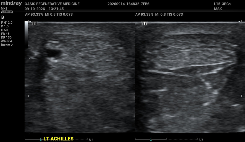
Above: the injured Achilles beside the healthy one on the same screen. The dark gap in the left-hand tendon is the tear, 2 mm across. Below: the same injured tendon four minutes later, on a probe built for structures this close to the skin, where the tear goes from a smudge to something you can measure. An X-ray shows none of this.

What we send elsewhere. A fracture picked up on ultrasound that an X-ray missed goes to a specialist because the foot bears weight, and that makes fracture management there less forgiving. We don't do wound care, so if a diabetic foot wound is a significant part of your presentation and nobody is managing it, we'll refer that part out. Ingrown and fungal toenails aren't a job for regenerative medicine.
How regenerative medicine compares to orthotics, cortisone, therapy, shockwave, and surgery
Regenerative medicine vs. orthotics. If the root cause was footwear with no meaningful arch support, correcting that is essential and sometimes most of the answer. The limitation is timing. By the time people reach us, the downstream problem is often self-sustaining, so you can correct the original cause and the tissue keeps misbehaving on its own.
Regenerative medicine vs. physical therapy and loading. Also essential, and also frequently not enough on its own. Tissue responds to load, and therapy restores proper force transmission and gets the stabilizing muscles doing their jobs. It stays part of the plan, regardless of what else we do.
Regenerative medicine vs. cortisone. Cortisone isn't evil; it's the wrong tool. It's an anti-inflammatory, and plantar fasciitis isn't an inflammatory problem. Worse, injecting a corticosteroid into a ligament or tendon weakens it, and a weakened plantar fascia or Achilles can rupture. You may have made the pain go away, but that's switching off the fire alarm while the building is still burning. The same caution applies to an ankle that still feels unstable after a sprain, and to peroneal tendinopathy, where cortisone gets reached for fairly indiscriminately.
Regenerative medicine vs. shockwave. A fair question, and the answer for now is that we don't offer it. The literature suggests it can relieve plantar fasciitis symptoms, and it's non-invasive, though it isn't comfortable and it runs six to twelve sessions spread over weeks. Those sessions are the reason it isn't on offer here. People reach us having already spent a year or two on incremental measures, and another slow burn isn't what they came for. That may change.
Regenerative medicine vs. surgery. For a far-gone plantar fascia, surgeons can release it, and that generally does relieve the pain. What's less advertised is how the results read at long follow-up. In a study of 74 patients, followed for an average of nearly five years after complete plantar fasciotomy, 73% were satisfied with their symptoms and 74% would have the procedure again, but 44.4% still reported swelling or tenderness in the foot, improved from before surgery for most of them, but not gone.¹ Add the ordinary surgical risks and the foot's higher susceptibility to infection, and it's a lot to accept for that result.
Achilles surgery is a different conversation. For a full-thickness rupture, surgical repair doesn't consistently outperform non-operative management in a CAM boot with progressively reduced heel lifts over six to eight weeks. That's surprising, and far too individual to settle on a web page. For partial-thickness injuries, non-operative management wins. Standard non-operative management involves no orthobiologic at all. A CAM boot plus a PRP injection is a different proposition from a CAM boot alone.
What non-surgical foot and ankle treatment actually involves
First, we work out where it came from. If a knee or hip problem has you walking badly, we need to know whether that caused the plantar fasciitis or the plantar fasciitis caused it. Often both are true by now, each making the other worse. Not every case has an identifiable cause, and sometimes it's bad luck, but the correctable ones get ruled out before anything else happens.
PRP is first line for most of what we see here, including foot and ankle arthritis, where the argument against steroids is the same as anywhere else: symptom relief bought with accelerated wear and tear.
The PRP we offer takes one injection rather than a series. We use a high-dose, highly concentrated preparation placed under live ultrasound guidance, paired with a structured rehab program. That last part is close to mandatory, because tissue only rebuilds its strength when it's loaded. We can't make anyone do physical therapy, and if your schedule won't allow it, we'll still treat you, but we'll be honest about what you're giving up.
Injection day. A plantar fascia injection hurts. There's no version of that sentence that's gentler and still true. We mitigate it with cold spray and a local anesthetic, using ropivacaine rather than lidocaine, because lidocaine can be toxic to PRP and would undercut the whole point. For the Achilles, we can use a sural nerve block. A medial and lateral plantar nerve block is available for the plantar fascia, and we don't reach for it by default because blocking those nerves blocks motor function along with sensation. You'd leave here with a foot that isn't working properly and a fall risk attached. If you don't tolerate pain well, we'll discuss it.
Afterward. Expect a pain flare for a few days and a limp. You can walk and drive, though you won't enjoy the driving. Impact is out for a while, meaning no running, cutting, or jumping, with a return to impact sports usually around six weeks, after therapy and once you've been cleared. Pain relief tends to arrive over the first few weeks and function over the following months, and by three months you know roughly 80% of what the final result will be. People keep gaining after that. It's also where the comparison with a steroid gets stark, because at three months a steroid is wearing off while PRP is still climbing.
Then we check the work. Every PRP treatment here includes a repeat examination. That's a standard part of the plan, rather than an add-on. The ultrasound is in the room for those visits, so if you want to see the tissue again and compare it against where you started, say so and we'll scan you. And if you aren't progressing the way we expected, we scan without being asked, to rule out a complication and to make sure the original diagnosis was right.
Where this is unlikely to help. If we never identify what caused it and nothing changes, it can come back. And PRP asks your body to do the work, so a body that isn't in a position to heal will limit the result. Uncontrolled diabetes, an untreated thyroid problem, smoking, sedentary living, and nutritional deficiencies all narrow what's possible. And an open wound on the foot will need to heal before we treat it.
Diabetes is one we can put a number on: Hemoglobin A1C reflects roughly your last three months of blood sugar control. Most surgeons won't operate above 8%; Dr. Kiok's cutoff is 7.5%, and he has grown more conservative about it rather than less, because everything works better when the metabolic picture is optimized. Above that number, we'll get it under control first, and since plantar fasciitis isn't time-sensitive, nothing is lost by spending three months doing it properly. That isn't a referral away: OASIS runs its own GLP-1 program, so it stays part of the same plan with the same physician.
Learn more about PRP.
Our consultation isn't your typical doctor's appointment
One hour with Dr. Kiok. $500, credited toward your treatment plan if you decide to move forward. You leave with:
-
A specific diagnosis, not a label.
-
A treatment plan, not a referral or “let’s follow up in six weeks.”
-
A clear answer on whether you’re a candidate for regenerative medicine.
Do you take insurance?
No, we’re a cash-pay clinic. We don’t take insurance because we don’t believe a coverage desk should shape your care. Treatment plans are made in person, with you and Dr. Kiok, on a timeline that fits your goals. No delays, no denials.
Learn more about why regenerative medicine isn't covered by insurance.
Outcomes vary by patient, condition, and stage of disease. Whether regenerative medicine is right for your foot or ankle is determined during the consultation, based on the diagnosis and your individual goals.
1. Gibbons R, Mackie KE, Beveridge T, Hince D, Ammon P. Evaluation of long-term outcomes following plantar fasciotomy. Foot & Ankle International. 2018;39(11):1312-1319.
Ready for an actual answer?
If you've been walking on a bad foot for a year or more, if the label you were given has never quite explained what you feel, or if you've been told the only thing left is to live with it, book a consultation.
If you're not sure whether you'd be a candidate, that's exactly what the consultation is set up to figure out.